|
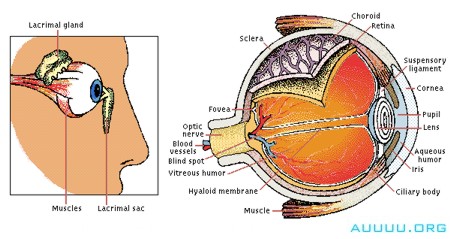
The amount of light entering the eye (right) is controlled by the pupil, which dilates and contracts accordingly. The cornea and lens, whose shape is adjusted by the ciliary body, focus the light on the retina, where receptors convert it into nerve signals that pass to the brain. A mesh of blood vessels, the choroid, supplies the retina with oxygen and sugar. Lacrimal glands (left) secrete tears that wash foreign bodies out of the eye and keep the cornea from drying out. Blinking compresses and releases the lacrimal sac, creating a suction that pulls excess moisture from the eye’s surface.
The entire eye, often called the eyeball, is a spherical structure approximately 2.5 cm (about 1 in) in diameter with a pronounced bulge on its forward surface. The outer part of the eye is composed of three layers of tissue. The outside layer is the sclera, a protective coating. It covers about five-sixths of the surface of the eye. At the front of the eyeball, it is continuous with the bulging, transparent cornea. The middle layer of the coating of the eye is the choroid, a vascular layer lining the posterior three-fifths of the eyeball. The choroid is continuous with the ciliary body and with the iris, which lies at the front of the eye. The innermost layer is the light-sensitive retina.
The cornea is a tough, five-layered membrane through which light is admitted to the interior of the eye. Behind the cornea is a chamber filled with clear, watery fluid, the aqueous humor, which separates the cornea from the crystalline lens. The lens itself is a flattened sphere constructed of a large number of transparent fibers arranged in layers. It is connected by ligaments to a ringlike muscle, called the ciliary muscle, which surrounds it. The ciliary muscle and its surrounding tissues form the ciliary body. This muscle, by flattening the lens or making it more nearly spherical, changes its focal length.
The pigmented iris hangs behind the cornea in front of the lens, and has a circular opening in its center. The size of its opening, the pupil, is controlled by a muscle around its edge. This muscle contracts or relaxes, making the pupil larger or smaller, to control the amount of light admitted to the eye.
Behind the lens the main body of the eye is filled with a transparent, jellylike substance, the vitreous humor, enclosed in a thin sac, the hyaloid membrane. The pressure of the vitreous humor keeps the eyeball distended.
The retina is a complex layer, composed largely of nerve cells. The light-sensitive receptor cells lie on the outer surface of the retina in front of a pigmented tissue layer. These cells take the form of rods or cones packed closely together like matches in a box. Directly behind the pupil is a small yellow-pigmented spot, the macula lutea, in the center of which is the fovea centralis, the area of greatest visual acuity of the eye. At the center of the fovea, the sensory layer is composed entirely of cone-shaped cells. Around the fovea both rod-shaped and cone-shaped cells are present, with the cone-shaped cells becoming fewer toward the periphery of the sensitive area. At the outer edges are only rod-shaped cells.
The eye is an organ. Behind its visible portions are a complicated array of delicate mechanisms that work in unison to transmit an image to the brain.
Where the optic nerve enters the eyeball, below and slightly to the inner side of the fovea, a small round area of the retina exists that has no light-sensitive cells. This optic disk forms the blind spot of the eye.
|